
Biomedical imaging
Overview of the computational imaging
Computational imaging describes a methodology that tailors the performance of an imaging system to advanced imaging and sensing tasks, by combing the steps of optical image formation and computational image processing. Figure 1 schematically illustrates the difference in the work flow between a conventional and a computational imaging system. A conventional imaging system incorporates an optical system to generate a direct image of an object scene, which is digitally recorded using an image sensor. A computational imaging system generates an optically encoded image using specialized optical hardware components. The digitally recorded image is subsequently decoded using computational processing. Optical encoding and digital decoding are linked by a mathematical model and optimized jointly in order to extract advanced object information of interest.

Fig.1 Overview of the computational imaging
Single-pixel imaging via spatial light modulation
As a strong competitor over conventional cameras featuring millions of pixels, a single-pixel camera images objects using structured illumination and synchronized intensity measurements with a bucket detector. Actually, it shifts the spatial information of objects onto a series of time-varying illumination patterns and computationally recovers the images with appropriate algorithms. In particular, the single-pixel cameras offer low-cost imaging systems beyond their pixelated counterparts at wavelengths or multiple wavelengths outside the visible spectrum, such as the infrared and X-ray range. Additionally, due to the superior detection efficiency and timing response of the single-pixel detector, such cameras enable imaging at weak intensity or time-resolved imaging with a high speed. Our lab combines the optical spatial modulation technology with the single pixel imaging by serving a series of complex-value optical fields as illumination beams. We develop several methods to achieve an extended depth of field with illumination via diffraction-free fields[1], isotropic edge detection for both amplitude[2] and phase objects and 3-D microscopic volumetric imaging[3].

FIG.2 Principle of the single-pixel phase imaging by complex Fourier spectrum sampling.
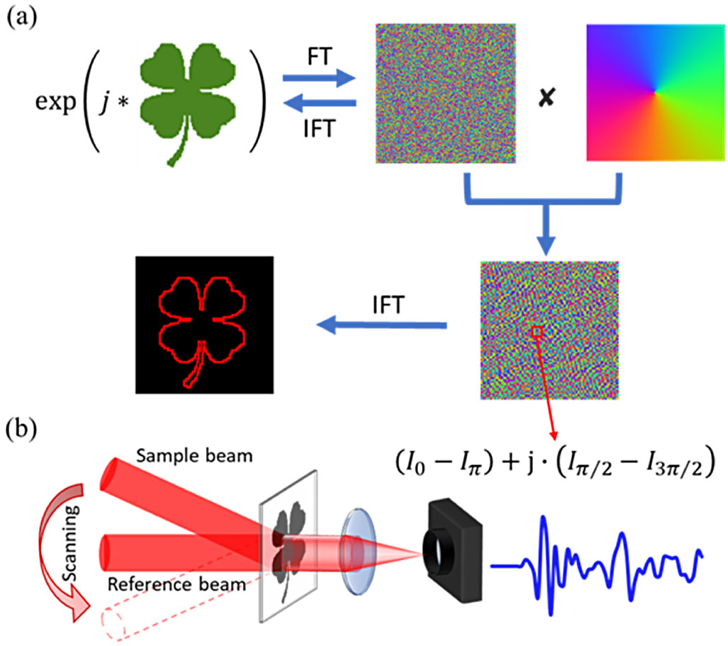
FIG.3 Principle of single-pixel spiral phase contrast imaging.
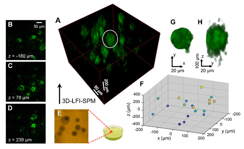
FIG.4 Label-free volumetric imaging of living Haematococcus pluvialis cells.
Photoacoustic microscopy
Photoacoustic microscopy is uniquely positioned for micro-biomedical imaging because of its ability to visualize optical absorption contrast in vivo directly and to facilitate either label-free or labeled 3D visualization of tissue. Based on photoacoustic effect, the photons of wavefront is converted into wide-band ultrasonic waves that can be detected using a high-frequency ultrasound transducer. Photoacoustic microscopy overcomes the strong scattering of light wave in biological tissues and is capable of achieving high-resolution images at greater depths than conventional microscopy methods. Here we propose motionless volumetric spatially invariant resolution photoacoustic microscopy, achieved through Fourier-spectrum optical excitation with simultaneous single-element photoacoustic detection at all depths. Our method achieves 1.5 times finer lateral resolution than conventional photoacoustic microscopy and is expected to open up new opportunities in biomedical applications. Figure 5 shows the depth-encoded whole-body images of a zebrafish larva obtained by our method (a) and conventional photoacoustic microscopy (b)[4].

FIG.5 Photoacoustic microscopy imaging of zebrafish embryos in vivo.
Lens-less endoscopic imaging via a single multimode fiber
Benefitting from the high capacity of transmitting various of orthogonal modes simultaneously and the minimally invasive observations in biomedical applications, the hair-thin multimode optical fiber is expected to be employed for endoscopic imaging. However, due to the dispersion and coupling of fiber modes, the light through multimode fiber is scrambled and mixed so that only the speckle pattern can be detected at the receiver. Recently, the wavefront shaping technology is introduced to obtain the precise knowledge of multimode fiber (namely, transmission matrix), which represents the linear relationship between the fiber input field and fiber output field. However, the interference-free experimental setup for calibrating transmission matrix and single-shot endoscopic imaging remains a challenge. Here, by combining wavefront modulation technology and deep learning technology, we propose a single-shot and lens-free endoscopic imaging method with a single multimode fiber to realize dynamic bioimaging in vivo without interference measurement[5].
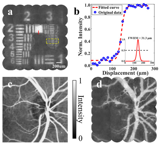
FIG.6 Endoscopic imaging with an enlarged field of view.
Edge Extraction by Spiral Phase Contrast Imaging
Image edge extraction enhances the contrast display of the image, which is a helpful approach for studying the structural characteristics of the imaging object. We propose a computational spiral phase contrast imaging method that utilizes symmetry-breaking spiral phase spatial filters under partially coherent illumination [Fig. 7(a)]. Such a method enables high image contrast and speckle suppression in edge detection simultaneously [Fig. 7(b)][6].

Fig. 6 Principle of spiral phase contrast imaging for edge extraction
Reference:
1.X. Hu, H. Zhang, Q. Zhao, P. Yu, Y. Li, and L. Gong, Single-pixel phase imaging by Fourier spectrum sampling, Applied Physics Letters 114, 051102 (2019).
2.Y. Liu, P. Yu, X. Hu, Z. Wang, Y. Li, and L. Gong, Single-pixel spiral phase contrast imaging, Optical Letter 45, 4028-4031 (2020).
3.Y. Liu, P. Yu, Y. Wu, J. Zhuang, Z. Wang, Y. Li, P. Lai, J. Liang, and L. Gong. Optical single-pixel volumetric imaging by three-dimensional light-field illumination. Proceedings of the National Academy of Sciences 120(31): e2304755120 (2023).
4.J. Yang, L. Gong, X. Xu, P. Hai, Y. Shen, Y. Suzuki, and L. V. Wang, Motionless volumetric photoacoustic microscopy with spatially invariant resolution, Nature Communications 8, 780 (2017).
5.Y. Liu, P. Yu, Y. Wu, Z. Wang, Y. Li, J. Liang, P. Lai and L. Gong. Single-shot wide-field imaging in reflection by using a single multimode fiber. Applied Physics Letters. 122, 063701 (2023).
6.S. Cheng, Y. Liu, P. Yu, Y. Wu, Z. Wang, Y. Li and L. Gong. Partially coherent spiral phase contrast imaging with a 3D-like effect. Applied Physics Express 15, 092004 (2022).